Smooth Muscle Diagrams : Microscopic Anatomy Of Muscle / These cells have fibers of actin and myosin which run through the cell and are supported by a framework of other proteins.
byAdmin•
0
Smooth Muscle Diagrams : Microscopic Anatomy Of Muscle / These cells have fibers of actin and myosin which run through the cell and are supported by a framework of other proteins.. They range from about 30 to 200 μm (thousands of times shorter than skeletal muscle fibers), and they produce their own connective tissue, endomysium.although they do not have striations and sarcomeres, smooth muscle fibers do have actin and myosin. In skeletal muscle, a single type of somatic nervous system traverses to muscle, where it stimulates organelle in the muscle cells in order to release calcium. Muscles are made up of highly specialized thin and elongated cells called muscle fibres.the muscle fibres contains specialized cytoplasm called sarcoplasm that contain network of the membrane called sarcoplasmic reticulum.the muscle fibres may be bounded by the cell membrane called sarcolemma.each muscle fibre may contain numerous. Smooth muscle anatomy smooth muscle tissue is also known as visceral muscle tissue. Related posts of smooth muscle diagram muscles of upper back.
Smooth muscle cells are found in the walls of hollow organs, including the stomach, intestines, urinary bladder and uterus, and in the walls of passageways, such as the. The smooth muscle of the alimentary canal (the digestive tract) facilitates the peristaltic waves that move swallowed food and nutrients. In this video i am gonna to show you how to draw the diagrams of cardiac, straited, smooth muscle for class 1st to 10th. Smooth muscles, cardiac muscles and skeletal muscles. There are three different types of muscles in the body:
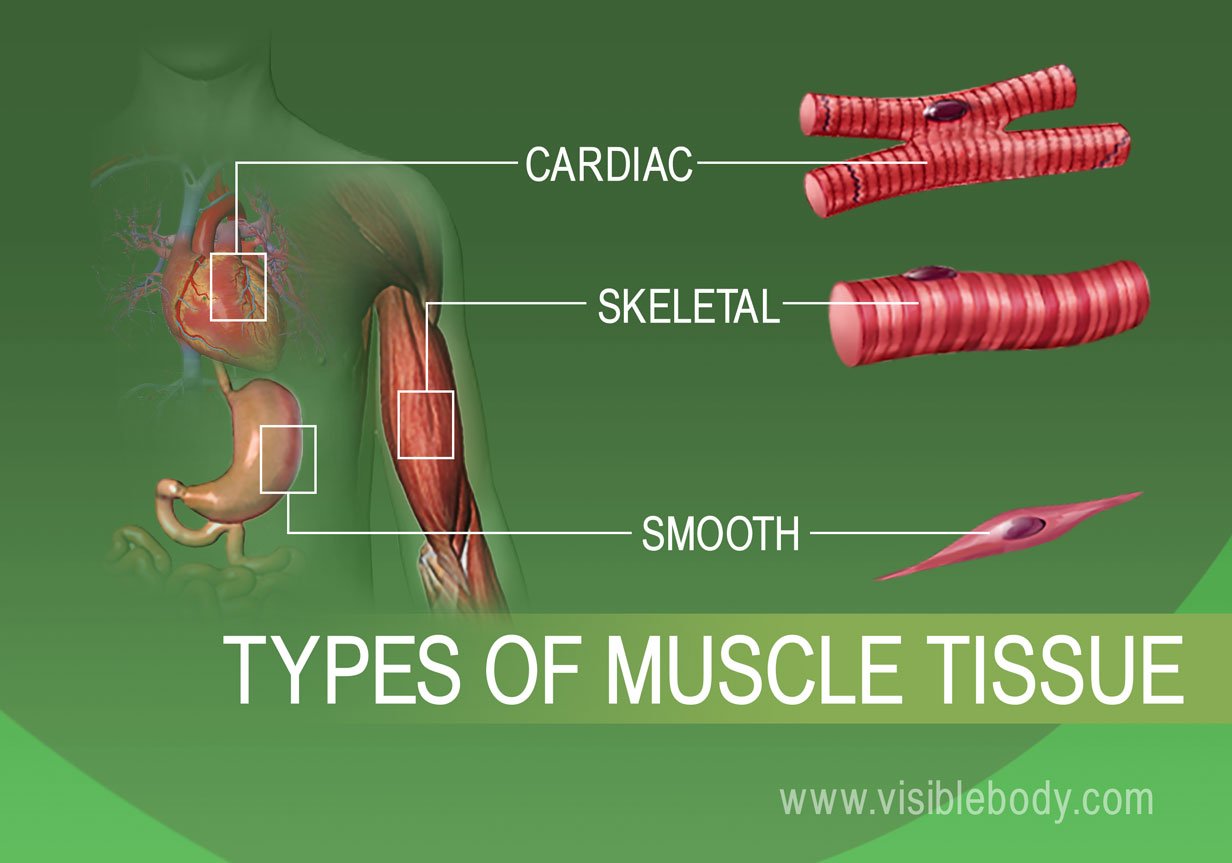
Muscle Tissue Types Learn Muscular Anatomy from www.visiblebody.com Smooth muscle contracts under certain stimuli as atp is freed. A body muscle diagram is used by different people for various uses. The three types of adrenoceptors present are: Related posts of smooth muscle diagram muscles of upper back. These cells have fibers of actin and myosin which run through the cell and are supported by a framework of other proteins. Muscles of upper back 12 photos of the muscles of upper back map of upper back muscles, muscles of the upper back and chest, origin and insertion of upper back muscles, superficial muscles of the upper back, tight muscles of the upper back and neck, human muscles, map of upper back muscles, muscles of the … Smooth muscle fibers are often found forming sheets of tissue and function in a coordinated fashion due to the presence of gap junctions between the cells. Smooth muscle (textus muscularis levis) smooth muscle is a type of tissue found in the walls of hollow organs, such as the intestines, uterus and stomach.
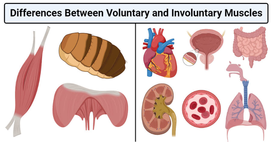
Smooth muscles are unique in their largely involuntary response, and in their structure.
Smooth muscles exhibits a phenomenon called _____ in which: Smooth muscle makes up the walls of hollow organs, respiratory passageways, and blood vessels. There are three types of muscles in the body: Smooth muscles, cardiac muscles and skeletal muscles. Smooth muscles have a much stronger ability to contract than skeletal. This smooth muscle can be found surrounding the walls of the blood vessels, the bronchioles in the lungs, and the sphincter muscles used in the gi tract.the gi tract, which is tubular by design, also houses longitudinal muscles in addition to the smooth. Related posts of smooth muscle diagram muscles of upper back. 101 diagrams free printable educational diagrams. It is found in numerous bodily systems, including the ophthalmic, reproductive, respiratory and gastrointestinal systems, where it functions to contract and cause movements under involuntary control. It is the pen diagram of skeletal, smooth and cardiac muscle for class 10, 11 and 12. It is layered in a distinctive pattern of circular layers. Muscles are made up of highly specialized thin and elongated cells called muscle fibres.the muscle fibres contains specialized cytoplasm called sarcoplasm that contain network of the membrane called sarcoplasmic reticulum.the muscle fibres may be bounded by the cell membrane called sarcolemma.each muscle fibre may contain numerous. • smooth muscles respond to stretch only briefly, and then adapts to its new length • the new length however, retains its original _____ seconds or minutes after it has been elongated or shortened (e.g.
Skeletal, smooth and cardiac muscle. There are three types of muscles in the body: Smooth muscle tissue, unlike striated muscle, contracts slowly and automatically. Some of these muscles are quite large and cover broad areas. Diagram of artery with smooth muscle identification understanding smooth muscles.
Voluntary Vs Involuntary Muscles Definition 16 Differences Examples from microbenotes.com Its wavelike movements propel things through the bodily system, such as food through. In skeletal muscle, a single type of somatic nervous system traverses to muscle, where it stimulates organelle in the muscle cells in order to release calcium. It constitutes much of the musculature of Muscles often contract to hold the body still or in a particular position rather than to cause. Termed unitary smooth muscle or visceral muscle, this type of smooth muscle is the most common observed in the human body, forming the walls of hollow organs. Muscles of upper back 12 photos of the muscles of upper back map of upper back muscles, muscles of the upper back and chest, origin and insertion of upper back muscles, superficial muscles of the upper back, tight muscles of the upper back and neck, human muscles, map of upper back muscles, muscles of the … Skeletal, smooth and cardiac muscle. Related posts of smooth muscle diagram muscles of upper back.
Certain back muscles extend to other areas, like the shoulders, upper arms, and thighs.
There are three types of muscles in the body: The smooth muscles perform the functions in the contrast of other types of muscles. Certain back muscles extend to other areas, like the shoulders, upper arms, and thighs. The heart muscle, smooth muscles, and skeletal muscles. In this video i am gonna to show you how to draw the diagrams of cardiac, straited, smooth muscle for class 1st to 10th. It is layered in a distinctive pattern of circular layers. Other muscles are small and cover much less space. Muscles of upper back 12 photos of the muscles of upper back map of upper back muscles, muscles of the upper back and chest, origin and insertion of upper back muscles, superficial muscles of the upper back, tight muscles of the upper back and neck, human muscles, map of upper back muscles, muscles of the … In skeletal muscle, a single type of somatic nervous system traverses to muscle, where it stimulates organelle in the muscle cells in order to release calcium. Its wavelike movements propel things through the bodily system, such as food through. In response to specific stimuli in smooth muscle, the intracellular concentration of ca 2+ increases, and this activator ca 2+ combines with the acidic protein calmodulin. Related to the function of movement is the muscular system's second function: It is the pen diagram of skeletal, smooth and cardiac muscle for class 10, 11 and 12.
Smooth muscle is composed of sheets or strands of smooth muscle cells. Smooth muscle tissue, unlike striated muscle, contracts slowly and automatically. Its wavelike movements propel things through the bodily system, such as food through. Smooth muscles have a much stronger ability to contract than skeletal. There are three types of muscles in the body:
Smooth Muscle Anatomy Mnemonic Contraction Multi Unit Vs Single Unit from www.registerednursern.com Muscles often contract to hold the body still or in a particular position rather than to cause. Smooth muscles have a much stronger ability to contract than skeletal. Related posts of smooth muscle diagram muscles of upper back. Vascular smooth muscle refers to the particular type of smooth muscle found within, and composing the majority of the wall of blood vessels. It is layered in a distinctive pattern of circular layers. It constitutes much of the musculature of Learn vocabulary, terms, and more with flashcards, games, and other study tools. Diagram of artery with smooth muscle identification understanding smooth muscles.
Related posts of smooth muscle labelled diagram muscle anatomy groin.
Some of these muscles are quite large and cover broad areas. Muscles often contract to hold the body still or in a particular position rather than to cause. In this video i have shown the simplest way of drawing muscle drawing. In response to specific stimuli in smooth muscle, the intracellular concentration of ca 2+ increases, and this activator ca 2+ combines with the acidic protein calmodulin. The maintenance of posture and body position. Related posts of smooth muscle diagram muscles of upper back. Smooth muscles exhibits a phenomenon called _____ in which: Smooth muscle makes up the walls of hollow organs, respiratory passageways, and blood vessels. The smooth muscles are often found within the organs and structures of organs. The three types of adrenoceptors present are: Your skeletal muscles are attached to your bones via tendons. There are three different types of muscles in the body: The smooth muscle in the walls of organs like the urinary bladder and the uterus allow those organs to expand and relax as needed.
Your skeletal muscles are attached to your bones via tendons smooth muscle diagram. Smooth muscles, cardiac muscles and skeletal muscles.